Posted by star
on 2019-06-05 19:35:37
Hits:1257
Adherens junctions and desmosomes are two types of adhesive junctions using homophilic (like to like) interactions of cadherins to bind epithelial cells to their neighbors. Cytoplasmic actin filaments reinforce adherens junctions, whereas cytoplasmic intermediate filaments anchor desmosomes.
Homophilic interactions between densely clustered E-cad-herins bind adjacent cells together at adherens junctions. β- Catenin and a related protein called plakoglobin bind the cytoplasmic domains of E-cadherin. ß-Catenin not only regulates gene expression when it enters the nucleus as part of the Wnt signaling pathway but also interacts with several Cytoskeletal and signaling proteins associated with adherens junctions. α-Catenin has been a candidate to connect cadherins to actin filaments, since it can bind both ß-catenin and actin filaments. However, these two interactions appear to be mutually exclusive, so the link between cadherins and actin is still under investigation.
Adherens junctions are the first connections that are established between developing sheets of epithelial cells. Contact begins when cadherins on the tips of filopodia engage partner cadherins of the same type on another cell. The contact spreads laterally as more cadherins are recruited along with associated actin filaments, as is illustrated by dorsal closure of the ectoderm by Drosophila embryos. These pioneering adherens junctions eventually allow like cells to associate in epithelial sheets and to influence the maturation of the epithelium. Adherens junctions are a prerequisite for the tight junctions that allow epithelial cells to establish polarity with different proteins and lipids in the apical and basal plasma membranes. The shape of the cells depends on Rho family GTPases and protein kinases associated with the adherens junction, which regulate the assembly and contraction of the associated actin cytoskeleton. The junctions and polarity of the cells determine t......
Posted by star
on 2019-06-05 18:51:36
Hits:249
In eukaryotic cells, the DNA carrying the genetic information is in the nucleus and the ribosomes that translate the genetic information are in the cytoplasm, they are separated by the nuclear membrane. RNA is the important mediation to cross nuclear membrane for genetic information. An RNA quality monitoring system is ubiquitous in the nucleus to rapidly identify and degrade mRNAs carrying erroneous genetic information or abnormalities, thereby ensuring accurate transmission of genetic information.
The highly conserved exosome complex is the most versatile RNA surveillance and degradation machine. Yet how it is negatively regulated remains clearly unknown.
Recently, researchers from China have discovered the first negative regulation factor NRDE2 of the RNA exosome complex. They clarified the functional mechanism how NRDE2 inhibits the RNA exosome complex and provided an important regulation mode of RNA nucleus export and degradation.
NRDE2 forms a 1:1 complex with MTR4(a nuclear exosome cofactor critical for exosome recruitment). The NRDE2 locates in nuclear speckles, where it inhibits MTR4 recruitment and RNA degradation to ensures efficient mRNA nuclear export.
The Structural and biochemical data revealed that NRDE2 interacts with MTR4’s key residues, locks MTR4 in a closed conformation, and inhibits MTR4 interaction with the exosome as well as proteins important for MTR4 recruitment, such as the cap-binding complex (CBC) and ZFC3H1. NRDE2 also inhibits the interaction between MTR4 and exosome, providing another important level of negative regulation. This NRDE2-mediated exosome negative regulation is critical for the dry maintenance of mouse embryonic stem cells.
The study first discovered the negative regulation factor NRDE2 of exosome , which explained its molecular mechanism and biological significance of inhibiting RNA degradation and promoting RNA nuclear transport, revealing a key mode regulating the nucleation or degrada......
Posted by star
on 2019-06-05 18:34:11
Hits:224
Tobacco use often produces precancerous cells that increase a person's risk of developing lung cancer, but researchers don't know how precancerous changes affect the development of cancer. Scientists from the duke university medical research center recently presented a new idea at the American association for cancer research, which suggests that these precancerous cells may turn nearby cells cancerous.
"We wanted to understand how these precancerous cells influence nearby cells to become cancerous," said researcher Dr. Kim a. Binder. "we studied the communication mechanism between precancerous lesions and cancer cells in the context of the presence of enzymes called PI3K.In most cancers, enzyme PI3K is activated, and the over-activation of PI3K is a necessary condition to drive carcinogenesis.PI3K is a kind of kinase, and there are many kinase inhibitors that can effectively resist the spread of cancer cells. Kinase inhibitors are effective against PI3K and do a good job of killing cancer cells grown in petri dishes, but they are not effective in patients' bodies, which is a noteworthy phenomenon.
The current study provides an important clue that cancer cell lines in petri dishes are very sensitive to PI3K inhibition, but in the vicinity of precancerous cells, these cancer cells become resistant to inhibitors. The researchers cultured lung cancer cells in the same petri dishes as precancerous cells, and then attacked the cells with PI3K inhibitors alone or together. The results showed that cancer cells that grew together with precancerous cells grew faster and developed tolerance to PI3K inhibitors than cancer cells that grew alone.
In addition, precancerous cells can stimulate the production of cancer stem cell-like properties in receptor cancer cells, which means that in addition to being able to resist PI3K inhibitor therapy, cancer cells next to precancerous cells may become more dangerous, such as the cancer cells spreading again. Or that canc......
Posted by star
on 2019-06-05 01:47:47
Hits:253
Scientists at duke university medical research center have identified a potential new target for regulating inflammation, which is linked to a range of diseases, including diabetes, cancer and alzheimer's. The potential target is an immune protein called SARM, which is very conserved throughout evolution and therefore very similar in humans and other mammals.
The innate immune system is activated as a protective mechanism, responding to the presence of pathogens or tissue damage. This innate immune activation leads to inflammation. However, excessive or inappropriate inflammation is associated with a range of debilitating diseases, so controlling inflammation is a major problem that scientists are trying to solve.
In general, inflammasomes are tiny molecular machines that assemble in immune cells after sensing infection or injury and activate the inflammatory response. When inflammatory corpuscles gather, they trigger the release of inflammatory cytokines, triggering inflammatory cell death.
But exactly what controls il-1 production and the extent to which apoptosis occurs during inflammation remains unclear. Experimental results show that SARM is a key regulatory factor of inflammatory body.
Scientists have shown that the more SARM cells contain, the less il-1 they produce, because SARM interferes with the assembly of inflammatory corpuscles. On the contrary, too much SARM can also cause cell death, because SARM causes damage to mitochondria.
Dr Johnstone, the researcher, said: "we've been trying to unravel the mystery of this immune protein - what exactly does it do at some point?"It may be a key regulator of inflammatory corpuscles, suggesting that SARM plays a role in inflammatory disease."
Scientists already know that SARM causes brain cell death, so it's being studied as a target for neurodegeneration and related diseases, and it's also being found to be a key regulator. The findings offer new hope that research into SARM, if p......
Posted by star
on 2019-06-04 18:59:50
Hits:266
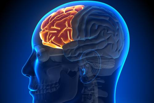
Neuroscientists from Boston University have found that age-related working memory impairments can be repaired. They found that this was possible by stimulating the brain by alternating- current stimulation.
The study involved 42 young people aged 20 to 29 and 42 elderly people aged 60 to 76. The researchers tested the performance of working memory activities in all subjects.
Researchers used the method of transcranial alternating current stimulation (TACS) to input electrical charge into the brains of young and old groups. They found that the use of variable current stimuli assisted 42 elderly people to restore normal electrogram rhythm in the left prefrontal cortex and left temporal cortex (these brain regions and working memory-related) . Without brain stimulation, the memory of older people is slower than that of young people, and the correct rate is lower too. This is because the specific brainwave frequencies of young people are highly correlated.
When older people are stimulated by positive brains, they can increase their working memory test scores to the level of young people, and the effect lasts for at least 50 minutes. When the elderly received 25 minutes electrical stimulation to their brains, the elderly are similar to those in their 20s who remember whether the on-screen image is the same or slightly changed from the previous version.
This suggests that treatment of such frequencies in the elderly's brain may help the brain function. The findings will provide the basis for more research and are expected to develop non-invasive tool to treat brain-complementing diseases such as schizophrenia, autism and Alzheimer's disease.